ORAL SURGERY

Wisdom Tooth Extractions: Surgical & Non-Surgical Impaction Care

Relieve painful impacted wisdom teeth, chronic gum swelling, and pressure on adjacent teeth. Safely planned with 3D CBCT imaging to protect mandibular nerves and performed under gentle anesthesia.

WhatsApp Us Call Us
20+ Years Trusted Care Digital 3D Accuracy Sarvodaya Enclave, South Delhi
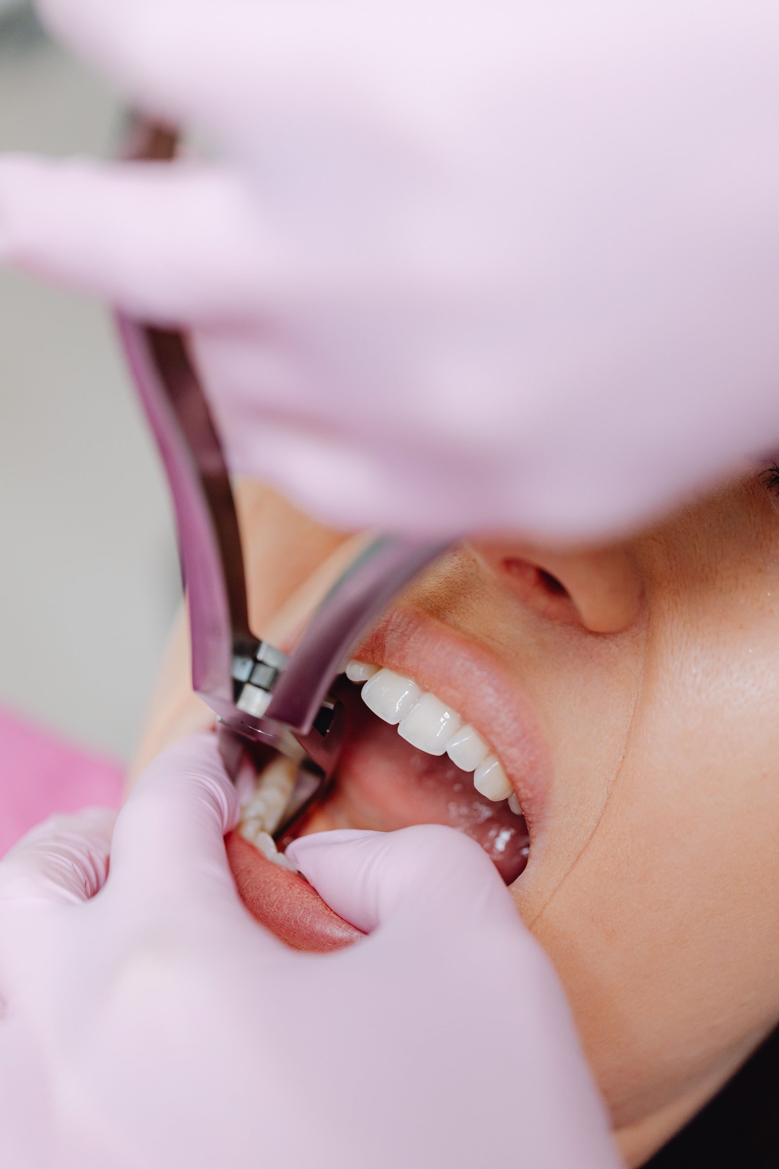
IMPACTION SPECIALIST Wisdom tooth extraction treatment
CBCT 3D Nerve MappingPiezo-Surgical Bone SparingRapid Post-Op Healing
AS
Dr. Amit Srivastava (BDS, MDS Orthodontics) 29+ Years Clinical Practice • Sarvodaya Enclave
Clinical Deep-Dive

Expert Management of Impacted Third Molars with 3D CBCT Safety

Wisdom teeth (third molars) are the last teeth to erupt, typically emerging between ages 17 and 25. In modern humans with smaller jaw dimensions, there is frequently insufficient space for them to emerge normally. As a result, wisdom teeth become impacted—tilted forward into adjacent molars (mesioangular), lying completely horizontal in bone, or trapped under painful, infected gum flaps (pericoronitis).

At Dental Specialities in Sarvodaya Enclave, we plan every surgical extraction using 3D CBCT scans. This allows Dr. Amit to map the exact 3D spatial relationship between the wisdom tooth roots and the inferior alveolar nerve canal, virtually eliminating the risk of nerve injury.

Our surgical technique is minimally invasive. Using delicate micro-sectioning, the tooth is divided into small, manageable pieces that glide out through a conservative bone window. This prevents bone trauma, drastically minimizes post-operative facial swelling, and ensures a smooth, rapid recovery.

Why Choose This Care

Key Benefits & Clinical Advantages

Permanent Pain & Infection Relief

Eliminates recurrent episodes of painful pericoronitis (infected gum flaps) and facial swelling.

Protects Adjacent Second Molars

Prevents food impaction and destructive root cavities on the critical neighboring chewing molars.

3D CBCT Nerve Mapping Safety

Pre-operative 3D imaging verifies exact nerve canal coordinates, planning anatomical safety.

Prevents Dentigerous Cysts

Eliminates the risk of fluid-filled cysts forming around unerupted crowns that can hollow out jawbone.

comfortable Surgical Execution

Profound, computer-guided local anesthesia ensures you feel zero sharp pain during surgery.

Minimally Invasive Sectioning

Tooth sectioning minimizes bone removal, leading to faster healing and minimal cheek swelling.

Candidate Suitability

Is This Treatment Right for You?

A comprehensive examination and digital scan confirms suitability. Here is our clinical guideline.

When This Treatment Is Recommended

  • Partially erupted wisdom teeth causing recurrent gum swelling, bad taste, and trismus
  • Horizontally or angularly impacted wisdom teeth pushing into the roots of adjacent molars
  • Wisdom teeth with deep dental decay or bone cysts that cannot be restored
  • Patients experiencing referred earaches, temple headaches, and jaw stiffness
  • Preventive removal in young adults aged 17–25 before root tips fully form and anchor

When An Alternative May Be Indicated

  • Fully erupted, upright wisdom teeth that are completely functional and easy to clean with a brush
  • Deep, completely bone-impacted wisdom teeth that are asymptomatic in elderly patients
How It Works

Step-by-Step Clinical Process

Transparent, predictable steps so you feel fully informed and confident at every stage.

01

3D CBCT Nerve Mapping Scan

Evaluating exact root angulation, bone depth, and the 3D course of the mandibular nerve.

02

Profound comfortable Local Numbing

Complete anesthesia of the surgical site so you feel relaxed and completely comfortable.

03

Micro-Surgical Gentle Sectioning

Creating a small conservative tissue window and gently sectioning the tooth for atraumatic removal.

04

Saline Cleansing & Resorbable Sutures

Cleansing the socket, placing resorbable dissolving sutures, and reviewing cold-pack post-op protocols.

How we plan your care

A clear process from consultation to review.

We use clinical assessment and digital planning to discuss the appropriate treatment—not patient result imagery.

1

Consult

We listen to your concerns and assess your oral health.

2

3D scan

iTero or CBCT imaging is used when it helps plan your care.

3

Digital plan

We explain appropriate options, stages and your treatment estimate.

4

Treatment & review

Care is monitored with planned check-ins and follow-up guidance.

Patient Inquiries

Frequently Asked Questions

Clear, clinically honest answers to the questions our patients ask most often.

Will I be put to sleep for wisdom tooth surgery?+

Most wisdom tooth extractions are performed smoothly and comfortably under modern local anesthesia. You are awake and completely comfortable, feeling only gentle pressure. For highly anxious patients, mild conscious sedation options are available.

How much swelling should I expect after surgery?+

Mild cheek swelling is a normal biological response that peaks around 48 hours after surgery and then resolves quickly. Applying an ice pack intermittently for the first 24 hours and taking prescribed anti-inflammatory medication keeps swelling minimal.

When can I return to work or college?+

Most patients have surgery on a Friday or Saturday and are back to work or classes by Monday. We advise avoiding vigorous gym workouts or heavy lifting for 3 to 4 days.

Do the stitches need to be removed?+

We typically place resorbable (dissolving) sutures that dissolve naturally on their own within 7 to 10 days, meaning you do not have to worry about suture removal.

Why is it better to remove wisdom teeth when young?+

Between ages 17 and 24, wisdom tooth roots are only one-third to two-thirds formed, surrounding bone is softer and more flexible, and the risk of nerve proximity is lowest. Younger patients also heal significantly faster.