
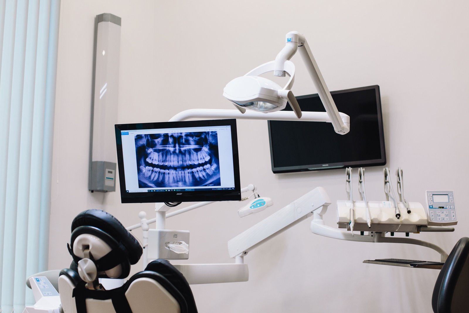
Traditional dental X-rays flatten three-dimensional human anatomy into a flat 2D picture. This results in superimposition, magnification distortion, and hides critical bone thickness details. Cone Beam Computed Tomography (CBCT) revolutionized dental diagnostics by capturing true 1:1 scale, isotropic 3D volumetric data.
Unlike medical CT scanners that expose patients to high radiation doses and require lying in an enclosed tunnel, dental CBCT uses a cone-shaped X-ray beam that orbits your head once while you sit or stand comfortably. It captures hundreds of planar projection views in under 20 seconds, using up to 95% less radiation than conventional hospital CT scanners.
At Dental Specialities in Sarvodaya Enclave, our CBCT scanner provides sub-millimeter slice resolutions (down to 75 microns). Dr. Amit slices, rotates, and navigates your jaw anatomy in axial, coronal, and sagittal cross-sections, allowing us to measure bone width to 0.1mm and map the exact path of the inferior alveolar nerve before performing implant surgery.