DENTAL RADIOLOGY

OPG Panoramic X-Ray in South Delhi: Comprehensive Full-Mouth Scan

Capture your entire mouth in a single seamless panoramic image. A fast, 12-second non-invasive scan that visualizes all 32 teeth, jawbones, wisdom tooth impactions, and TMJ joints with zero items placed inside your mouth.

WhatsApp Us Call Us
20+ Years Trusted Care Digital 3D Accuracy Sarvodaya Enclave, South Delhi
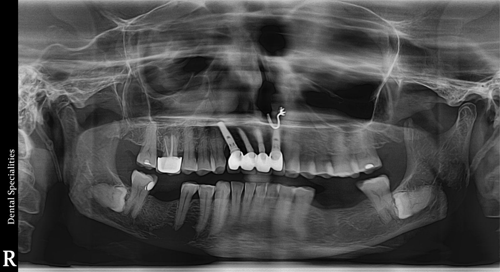
PANORAMIC RADIOLOGY Panoramic OPG dental X-ray
Full Panoramic Dentition ViewTMJ & Maxillary Sinus MappingQuick 12-Second Non-Invasive Scan
AS
Dr. Amit Srivastava (BDS, MDS Orthodontics) 29+ Years Clinical Practice • Sarvodaya Enclave
Clinical Deep-Dive

The Big Picture: Panoramic Imaging of Teeth, Jaws & TMJ Joints

An Orthopantomogram (OPG)—commonly called a panoramic dental X-ray—is an indispensable diagnostic tool that captures an expansive, wide-angle view of your entire lower face. Unlike small intraoral X-rays that focus on just two or three teeth, an OPG displays all upper and lower teeth, the maxillary sinuses, the advanced nickel-titaniuml cavity, and both temporomandibular (TMJ) jaw joints in a single continuous radiograph.

The scan is completely non-invasive and extraoral: no sensors or film packets are placed inside your mouth, making it exceptionally comfortable for patients with a sensitive gag reflex, children, or those experiencing lockjaw.

You simply stand in front of our modern digital panoramic unit while the rotating gantry glides smoothly around your head in just 12 to 15 seconds. The resulting digital image gives Dr. Amit a complete anatomical overview to plan orthodontics, evaluate unerupted wisdom teeth, and assess bone levels for full-mouth restorations.

Why Choose This Care

Key Benefits & Clinical Advantages

Entire Mouth in One Radiograph

Visualizes all 32 teeth, unerupted tooth buds, upper and lower jawbones, and sinuses simultaneously.

100% Non-Invasive Extraoral Scan

Nothing is placed inside your mouth; perfect for patients who gag easily or have difficulty opening wide.

Fast 12-Second Exposure

The rotating sensor captures the full panoramic scan in just seconds while you stand comfortably.

Essential for Wisdom Teeth & Braces

Accurately reveals whether wisdom teeth are horizontal or tilted and maps dental arch symmetry.

Bilateral TMJ Joint Evaluation

Compares both left and right condyle heads to diagnose jaw joint wear, arthritis, and dislocation.

Instant Digital Reporting

High-resolution digital image is available immediately for chairside consultation or digital export.

Candidate Suitability

Is This Treatment Right for You?

A comprehensive examination and digital scan confirms suitability. Here is our clinical guideline.

When This Treatment Is Recommended

  • Pre-orthodontic assessment before starting metal braces or Invisalign clear aligners
  • Evaluating the angulation and depth of impacted wisdom teeth before surgery
  • Patients with jaw joint clicking, popping, or locked jaw (TMJ disorders)
  • Screening for hidden jaw cysts, tumors, or unerupted supernumerary teeth
  • Full mouth dental implant planning and bone resorption evaluation

When An Alternative May Be Indicated

  • Detecting tiny hairline enamel cracks or microscopic interproximal decay (Small RVG X-ray preferred)
  • Complex dental implant surgeries requiring 3D cross-sectional slice measurements (3D CBCT Scan indicated)
How It Works

Step-by-Step Clinical Process

Transparent, predictable steps so you feel fully informed and confident at every stage.

01

Metal & Jewelry Preparation

Removing earrings, necklaces, hairpins, and glasses to ensure a crystal-clear artifact-free scan.

02

Comfortable Laser-Guided Positioning

Resting your chin on the sterile support while soft laser light beams verify exact anatomical alignment.

03

12-Second Panoramic Rotation

Standing completely still as the camera arm glides quietly around your head for 12 seconds.

04

Instant High-Def Image Review

Viewing the comprehensive panoramic radiograph on our HD monitors with Dr. Amit.

How we plan your care

A clear process from consultation to review.

We use clinical assessment and digital planning to discuss the appropriate treatment—not patient result imagery.

1

Consult

We listen to your concerns and assess your oral health.

2

3D scan

iTero or CBCT imaging is used when it helps plan your care.

3

Digital plan

We explain appropriate options, stages and your treatment estimate.

4

Treatment & review

Care is monitored with planned check-ins and follow-up guidance.

Patient Inquiries

Frequently Asked Questions

Clear, clinically honest answers to the questions our patients ask most often.

Is an OPG X-ray uncomfortable?+

Not at all! You simply stand still and bite lightly on a small disposable plastic notch. The machine moves around your head without touching you. It is completely comfortable and stress-free.

How does an OPG compare to small dental X-rays?+

Small X-rays (intraoral RVGs) give high magnification of 2-3 individual teeth to check for small cavities. An OPG gives the broad 'panoramic landscape' of your entire jaw, wisdom teeth, sinuses, and TMJ joints.

Can I get an OPG if I gag easily?+

Yes! Because the OPG is completely extraoral (nothing is inserted into the back of your mouth), patients who gag with regular dental film experience zero gagging with an OPG.

What is the radiation dose of a digital OPG?+

Our digital OPG machine utilizes ultra-low radiation protocols. The dose is roughly equivalent to a 2-hour commercial flight, making it safe for adults and children alike.

Can I receive the digital OPG file to take to another specialist?+

Yes, we can instantly export your high-resolution digital OPG file in standard JPEG or DICOM format and send it via email or WhatsApp.