DENTAL RADIOLOGY

Intraoral X-Ray (RVG): Ultra-Low Radiation Instant Digital Diagnostics

High-definition digital radiography that reveals hidden cavities between teeth, bone levels, and root infections in seconds. Engineered with advanced CMOS sensors delivering 90% less radiation than traditional film.

WhatsApp Us Call Us
20+ Years Trusted Care Digital 3D Accuracy Sarvodaya Enclave, South Delhi
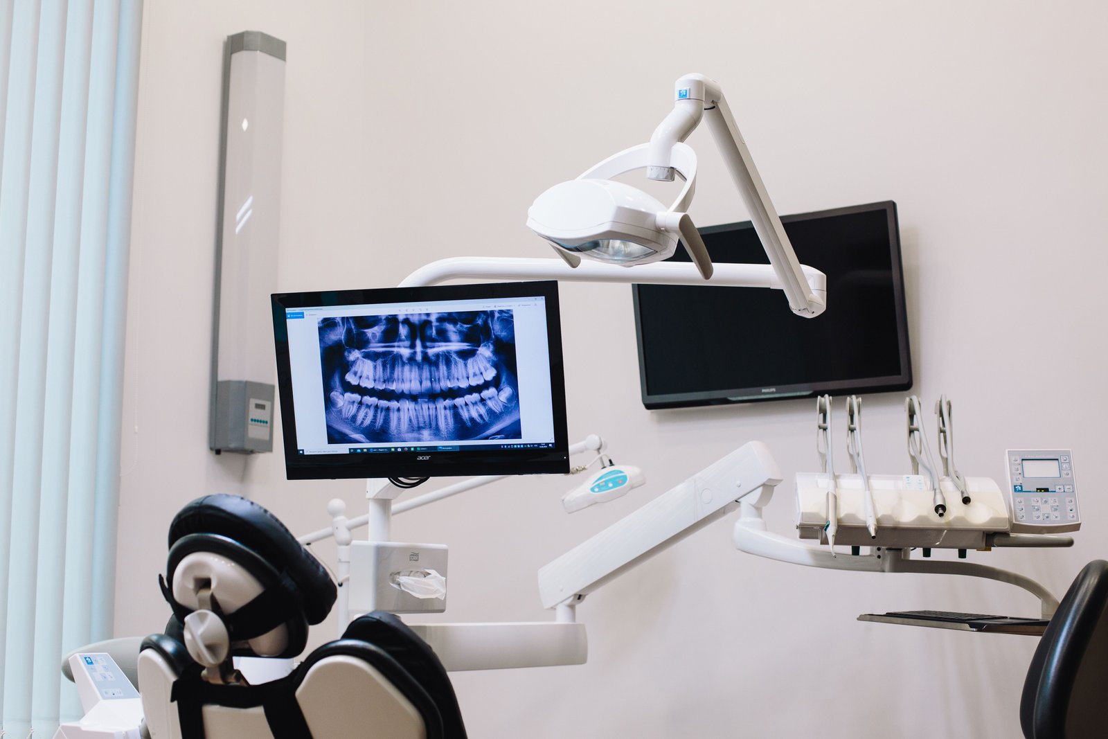
DIGITAL RADIOLOGY Dental radiography equipment in use
HD CMOS Sensor Resolution90% Lower Dose than FilmInstant Chairside Imaging
AS
Dr. Amit Srivastava (BDS, MDS Orthodontics) 29+ Years Clinical Practice • Sarvodaya Enclave
Clinical Deep-Dive

Sub-Millimeter Diagnostic Accuracy with 90% Lower Radiation

Over 40% of all dental conditions—including interproximal cavities between teeth, early bone loss, internal tooth resorption, and root tip abscesses—are completely invisible to the naked human eye during a routine mirror examination. High-resolution digital intraoral radiography (RVG) makes the invisible visible.

At Dental Specialities in Sarvodaya Enclave, we use high-sensitivity CMOS digital sensors with ergonomic rounded corners for total patient comfort. Unlike old analog film that required toxic chemical developing and high radiation doses, digital RVG captures crystalline radiographs instantly on our chairside monitors with up to 90% lower radiation exposure.

Our diagnostic software allows Dr. Amit to zoom in, enhance contrast, measure canal lengths to a fraction of a millimeter, and walk you through your scan on an HD screen so you understand your dental health with complete transparency.

Why Choose This Care

Key Benefits & Clinical Advantages

90% Less Radiation Than Film

Digital sensors require only a fraction of a second of exposure, making it exceptionally safe for adults and children.

Instant Chairside Results

Image appears on the HD screen in 3 seconds, eliminating long waits for chemical darkroom developing.

Detects Hidden Cavities Early

Reveals microscopic decay between tight teeth long before it causes pain or requires a root canal.

Sub-Millimeter Root & Bone Clarity

High-contrast resolution visualizes root canal anatomy, bone loss, and periapical infections precisely.

Transparent Patient Education

You view your X-ray directly on screen alongside Dr. Amit, making diagnosis completely clear and transparent.

Eco-Friendly & Non-Toxic

100% digital technology completely eliminates hazardous lead foil and toxic developing chemicals.

Candidate Suitability

Is This Treatment Right for You?

A comprehensive examination and digital scan confirms suitability. Here is our clinical guideline.

When This Treatment Is Recommended

  • Patients experiencing sudden toothache, temperature sensitivity, or pain on biting
  • Routine 12-month checkup to check for hidden decay between teeth
  • Root canal therapy monitoring to verify canal lengths and hermetic sealing
  • Post-trauma evaluation following sports impact or accidental falls
  • Evaluating bone levels around existing dental implants

When An Alternative May Be Indicated

  • Full-mouth comprehensive survey of jaw joints, wisdom teeth, and sinuses (Panoramic OPG X-ray indicated)
  • 3D bone volume mapping for dental implants or complex impactions (3D CBCT Scan indicated)
How It Works

Step-by-Step Clinical Process

Transparent, predictable steps so you feel fully informed and confident at every stage.

01

Ergonomic Sensor Placement

Placing the cushioned, rounded digital sensor gently inside the mouth adjacent to the target tooth.

02

Low-Dose Micro-Exposure

A fraction-of-a-second micro-exposure from our shielded, high-frequency DC X-ray tube.

03

Instant Image Transfer

The high-definition radiograph appears instantaneously on the dental chair monitor.

04

Diagnostic Review & Explanation

Dr. Amit magnifies the image, points out findings clearly, and formulates an honest, tailored plan.

How we plan your care

A clear process from consultation to review.

We use clinical assessment and digital planning to discuss the appropriate treatment—not patient result imagery.

1

Consult

We listen to your concerns and assess your oral health.

2

3D scan

iTero or CBCT imaging is used when it helps plan your care.

3

Digital plan

We explain appropriate options, stages and your treatment estimate.

4

Treatment & review

Care is monitored with planned check-ins and follow-up guidance.

Patient Inquiries

Frequently Asked Questions

Clear, clinically honest answers to the questions our patients ask most often.

How safe is a digital dental X-ray?+

Extremely safe! A single digital RVG X-ray exposes you to approximately 0.005 mSv of radiation—equivalent to the natural background radiation you receive simply by spending 1 to 2 hours outside in normal sunlight.

Can pregnant women have dental X-rays?+

Routine elective dental X-rays are typically postponed during pregnancy. However, if there is a severe dental emergency, infection, or pain, a digital X-ray can be safely taken with our lead apron and thyroid collar shield.

Will the sensor hurt my mouth or make me gag?+

Our digital sensors feature soft, beveled edges and ergonomic positioning holders specifically engineered to prevent palate poking and eliminate the gag reflex.

Can I receive a digital copy of my X-ray?+

Yes! Because all our radiographs are stored digitally in your electronic health record, we can email or WhatsApp your high-resolution image directly to you at any time.

Why do I need an X-ray if my tooth doesn't hurt?+

Dental decay and bone infections often develop silently for months or years without causing any pain until they reach the inner nerve. Early detection on digital X-rays saves you from expensive root canals or tooth loss.